Picture a traditional microscope transformed into a high-tech powerhouse. That's what a whole slide scanner accomplishes – combining precise robotics, sophisticated computer control, and cutting-edge optical sensors to capture tissue samples in unprecedented detail. This technological marvel serves as the foundation of modern digital pathology.
Scanning Modalities
Modern scanning employs two primary approaches: line scanning sweeps across slides in continuous motion, while tile scanning captures a grid of individual images. Both require “stitching” algorithms to create seamless whole-slide images.
To combat the eternal enemy of microscopy – poor focus – manufacturers deploy ingenious solutions. Dual-camera systems with a dedicated focusing camera work in tandem with the imaging sensor. Strobe lighting eliminates motion blur, while smart autofocus systems detect and rescan problematic areas automatically.

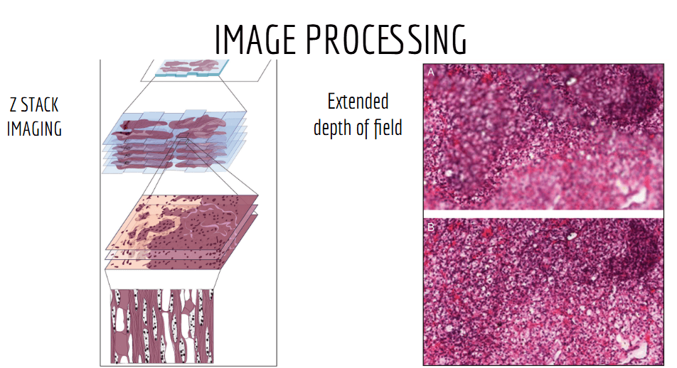
Once the scanning is complete, the digital images undergo a series of processing steps. This can include adjustments to brightness, contrast, and color to ensure the scanned image is a clear and accurate representation of the tissue sample. One particularly important technique in WSI is Z-stack imaging, which captures multiple focal planes at different depths along the z-axis. The images from these different planes are then combined to create a single, comprehensive image. This method is especially useful for thick tissue samples, where depth information is critical for accurate analysis.
However, technical challenges remain. Scanner artifacts can manifest as striping, stitching misalignments, or focus variations. Understanding these potential issues proves crucial for quality control and accurate diagnosis. Modern systems employ sophisticated detection algorithms to identify and correct many artifacts automatically.
Another major challenge of WSIs is the sheer size of the resulting image files. A single slide at 40X magnification generates around 16 billion pixels. That's equivalent to about 8,000 high-resolution smartphone photos in one image!
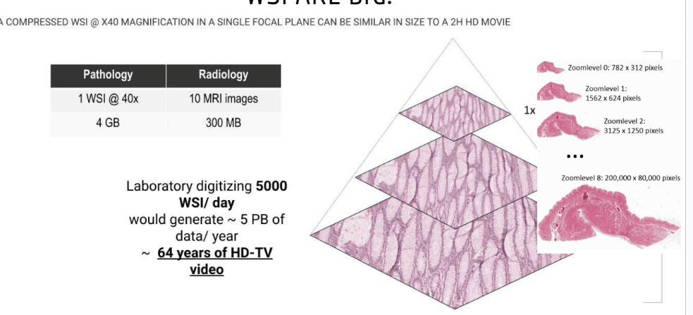
To manage this, scanners use data compression techniques and cloud storage systems. Images are typically stored at lower resolutions, with high-resolution data being retrieved only when the user zooms in on specific regions of interest. However, even compressed WSI files can still be quite large, often reaching around one gigabyte per slide.

Transforming workflow
Unlike traditional microscopy, where slides must be physically handled and examined, WSI allows for digital storage. Slides move directly from preparation to scanning, then secure storage on digital servers. Pathologists access these high-resolution images from anywhere within their secure network, eliminating the physical logistics of slide management. This transformation slashes turnaround times and enables unprecedented collaboration.
Despite the clear benefits, the adoption of whole slide imaging does come with its challenges. The initial cost of implementing WSI systems can be high, and there are ongoing concerns about the need for secure data storage, regulatory compliance, and the necessity for ongoing training. However, as technology advances and becomes more affordable, these challenges are gradually being addressed, enabling broader adoption across the medical field.
The Global Market Landscape
The WSI market features fierce competition among established players and innovative startups. Companies like Leica Biosystems, Roche (Ventana), Philips, and Hamamatsu dominate with comprehensive solutions. Each brings unique strengths: some excel in automation, others in image quality or software integration. This competitive landscape drives continuous innovation, promising even more capable systems in the future.
Continue reading…