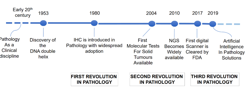
The story of pathology unfolds across three revolutionary waves. In the 19th century, the microscope transformed medicine by revealing the cellular universe, establishing the foundation of modern pathology. The second wave crashed through with molecular diagnostics, empowering pathologists to quantify biomarkers and peer into the genetic machinery of disease. Now, artificial intelligence surges forward as the third revolutionary force, amplifying human expertise with computational power.
AI’s application in digital pathology is deeply intertwined with advances in technology, particularly digital imaging. As pathology shifts from glass slides to digitized images, AI algorithms can extract critical information from these images much faster than a human can. These algorithms rely on vast quantities of data, including annotated images, to learn how to spot patterns and abnormalities that may go unnoticed by the human eye. This integration of AI is not just about increasing speed, but improving diagnostic accuracy, augmenting human expertise with a level of precision that could take years for a pathologist to accumulate.
A paradigm shift
Traditional image analysis relied on human-designed algorithms – experts painstakingly programmed specific features to detect. Machine learning revolutionized this approach. Instead of hard-coding rules, deep learning systems discover patterns from annotated examples. Imagine teaching a child to recognize cats – rather than listing every possible feature, you show them thousands of cat pictures. Similarly, modern AI systems learn from vast collections of pathology images, developing their own sophisticated pattern recognition abilities.
Pathology Informatics
One of the key challenges in the adoption of AI in digital pathology is ensuring that these models are transparent and understandable. Many AI systems are often referred to as "black boxes" because it can be difficult to interpret how they arrive at their conclusions. Pathology informatics seeks to address this by providing tools and frameworks that make AI’s decision-making process more transparent. The goal is to ensure that pathologists can trust AI-generated insights and understand the reasoning behind them, which is crucial for clinical adoption. As AI models become more interpretable, their use in pathology will grow, as both researchers and clinicians will have greater confidence in their results.
Areas where AI is making strides in pathology:
Ensuring High-Quality Digital Slides: Before any diagnosis, image quality matters supremely. AI serves as a vigilant guardian, detecting artifacts like air bubbles, tissue folds, or staining irregularities. These algorithms flag problematic slides for review, ensuring only pristine images advance to diagnosis. This automated quality control accelerates workflow while maintaining rigorous standards.
Disease detection: AI's most profound impact emerges in disease detection. While human pathologists excel at complex pattern recognition, AI never tires and can rapidly pre-screen thousands of images. The technology excels at tasks like detecting metastatic breast cancer in lymph nodes or grading prostate cancer severity. Rather than replacing pathologists, AI amplifies their capabilities, flagging suspicious regions for expert review.
Creating to learn: GANs represent one of AI's most fascinating innovations. These systems pit two neural networks against each other – one generating fake pathology images, the other detecting the fakes. Through this digital duel, GANs learn to create incredibly realistic synthetic images. This technology promises to solve critical challenges in pathology, from expanding limited datasets to creating standardized teaching materials.
PanProfiler: AI in Clinical Practice
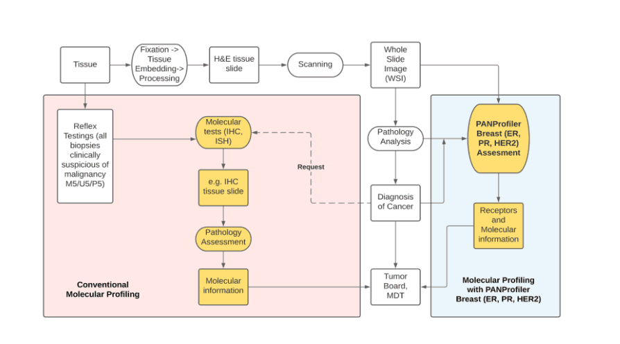
A prime example of AI’s impact on specific clinical challenges is PanProfiler, a system designed to analyze breast cancer markers like estrogen receptor (ER), progesterone receptor (PR), and HER2. PanProfiler demonstrates how AI and digital pathology work together to improve the accuracy of cancer diagnosis and treatment planning. By automating the detection and quantification of these markers, AI can significantly reduce the time required for analysis and provide more consistent results. This is particularly valuable in breast cancer treatment, where decisions regarding hormone therapy and targeted therapies are made based on these markers.
In traditional molecular profiling, pathologists rely on manual techniques to assess markers like ER, PR, and HER2, which can be time-consuming and prone to human error. PanProfiler, powered by AI, streamlines this process by automatically analyzing these markers, offering faster and more reliable results. This not only improves diagnostic efficiency but also ensures that treatment decisions are based on the most accurate and up-to-date information. AI systems like PanProfiler are pushing the boundaries of what’s possible in personalized medicine, particularly in oncology.
A new standard
Modern pathology laboratories increasingly embrace end-to-end AI integration. The workflow begins with automated scanning quality control, progresses through AI-powered triage that prioritizes urgent cases, provides real-time guidance during review, and concludes with comprehensive quality assurance. This seamless integration enhances efficiency while maintaining diagnostic accuracy.
Continue reading…