When you visit your doctor with concerning symptoms, they begin their investigation. Blood tests might tell part of the story, but sometimes it’s not enough. That's when radiology steps in – MRI and CT scans reveal a bigger picture. But, even these tools can't zoom in to the cellular level.
What if you're dealing with a persistent cough? Is it pneumonia or lung cancer? While radiologists spot suspicious growths, they can't always determine if they're benign or malignant. That's where pathology unleashes its power. Through biopsies, pathologists determine what’s wrong and how to treat it.
The journey of a tissue sample
When a surgeon performs an incisional biopsy, they remove a small piece of tissue to examine it for disease patterns. This is a meticulous process:
Fixation: The tissue is preserved, typically with formalin, to maintain its structure.
Embedding: After dehydration and clearing, the tissue is embedded in paraffin wax, creating a stable block for slicing.
Microtomy: A microtome cuts thin tissue sections from the wax block, making them ready for staining.
The art of staining
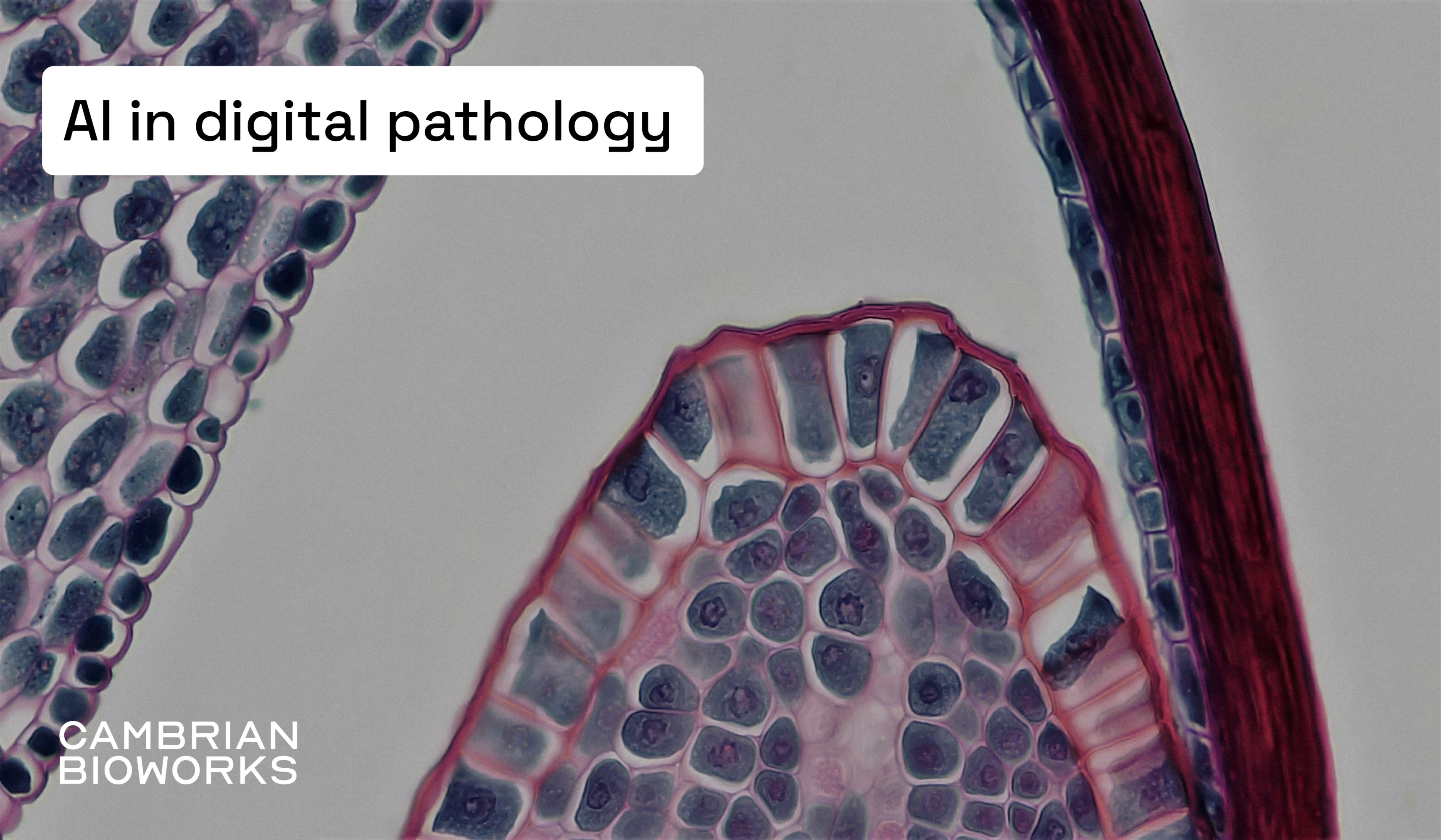
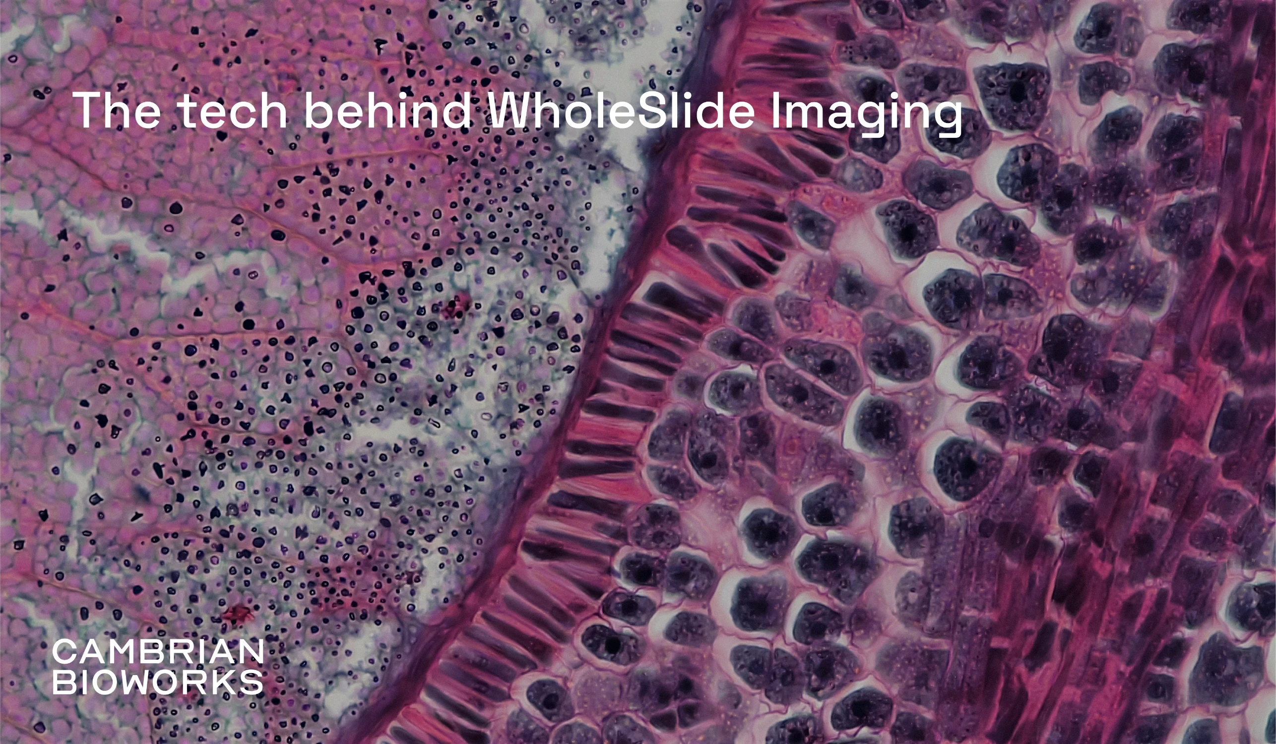
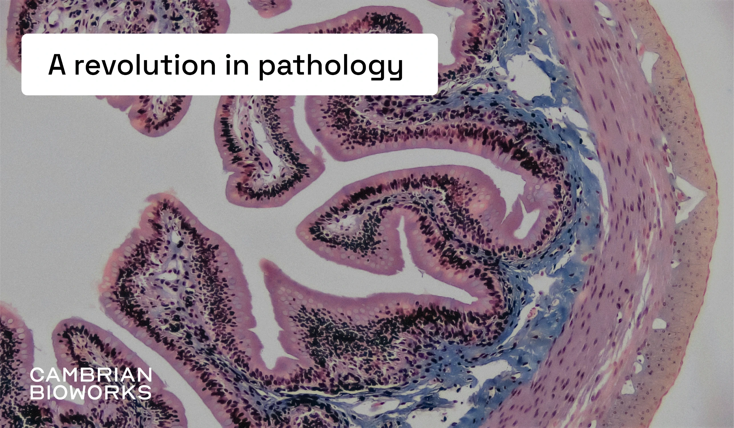
The world's gold standard for pathology is the hematoxylin and eosin (H&E) stain. This dynamic duo transforms transparent tissue into a vivid landscape of purples and pinks, revealing intricate cellular details. Hematoxylin is the purple dye that binds to acidic structures like DNA and RNA, staining the nuclei of cells. Eosin is the pink-orange dye that binds to basic structures like proteins in the cytoplasm and extracellular matrix. This provides a clear and detailed view of the tissue's structure and cellular details, helping pathologists identify abnormalities.
H&E staining dominates pathology labs worldwide because it's affordable, reliable, and consistently delivers excellent results.
Traditional pathology
While medical technology races forward, pathology labs seem stuck in a time capsule. For decades, pathologists have peered through microscopes, examining slides one by one. This traditional workflow moves like this:
The hospital sends samples to the pathology lab
Lab technicians prepare slides
Staff manually sort and distribute slides to pathologists
Pathologists examine and diagnose
Results return to the lab for archiving
This system has notable limitations. Physical slides and samples degrade over time, making long-term storage challenging. Diagnosis relies on manual sorting, which can be time-consuming and prone to errors. Collaboration is also restricted—obtaining a second opinion often means shipping slides or relying on tools not optimized for pathology.
The future of diagnostics
The future of diagnostics is here, and it’s digital. Digital pathology shatters old limitations, offering a whole new world of possibilities:
Immediate Expert Collaboration
Need a second opinion from a specialist across the country? Digital pathology enables instant slide-sharing and real-time consultations.
Enhanced Tumor Board Meetings
Medical teams can now examine digital slides together, zooming in on specific areas while discussing treatment strategies.
Powerful Analysis Tools
Precise annotations highlight areas of interest
Advanced image adjustment reveals subtle details
Quick retrieval of historical cases for comparison
Temporal data tracking shows disease progression
The digitalization of pathology marks a pivotal moment in medical history. By breaking free from physical constraints, we're entering an era of enhanced collaboration, more accurate diagnoses, and better patient outcomes. The microscope that served us for centuries now passes the torch to digital technology, for more efficient and accurate healthcare.
Continue reading…